42 spinal cord model with labels
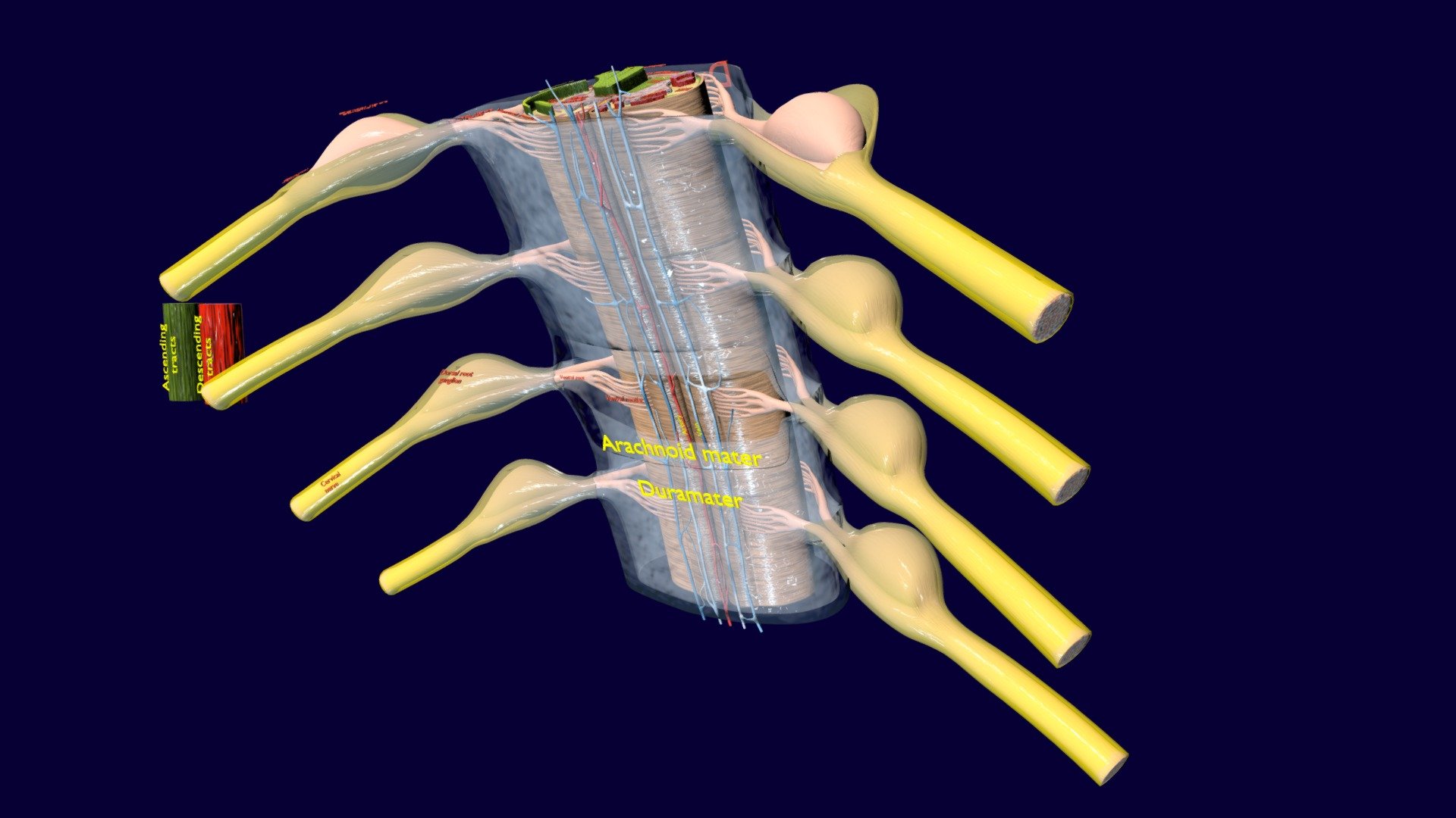
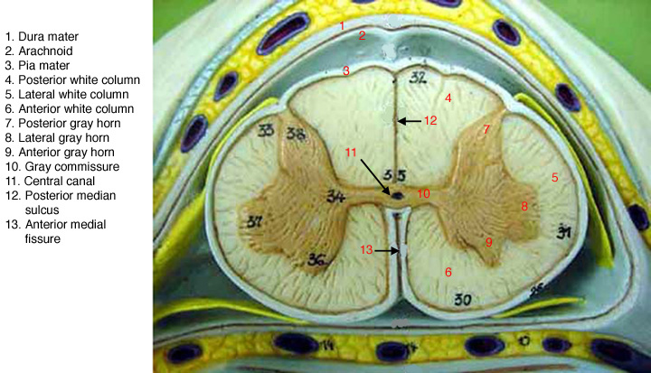
Solved Identify the following structures on the spinal cord - Chegg Identify the following structures on the spinal cord model & label the images below. Match the structures with the correct location using lecture notes Show transcribed image text Expert Answer 1.Duramater of spinal cordA 2. Arachnoid mater of spinal cord 3. Piamater of spinal cord 4. 5. 6. White matter 7. Dorsal horn 8. Lateral horn 9. The FASEB Journal - Wiley Online Library We are delighted to welcome Dr. Jeannine Botos to FASEB in the newly created role of Senior Managing Editor for The FASEB Journal and FASEB BioAdvances. Jeannine received a B.S. in Biochemistry and a Ph.D. in Veterinary Physiology and Pharmacology from Texas A&M and completed a postdoc at the National Cancer Institute.
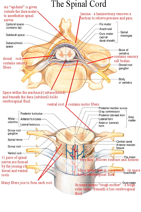
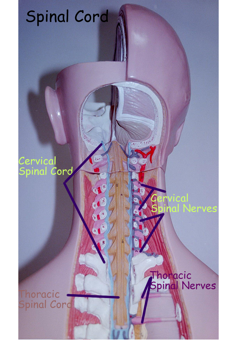
Spinal Cord - Anatomy, Structure, Function, & Diagram - BYJUS In adults, the spinal cord is usually 40cm long and 2cm wide. It forms a vital link between the brain and the body. The spinal cord is divided into five different parts. Sacral cord Lumbar cord Thoracic cord Cervical cord Coccygeal Several spinal nerves emerge out of each segment of the spinal cord.
Spinal cord model with labels
Spinal Cord Injury | Types of Spinal Cord Injuries ... 88 percent of spinal cord injury survivors who were single at the time of the accident are single five years later, compared to 65 percent in the general population. Two-thirds of sports-related spinal cord injuries are from diving, making it the most dangerous sport for the brain and spinal cord. Duke Neurosciences - Lab 2: Spinal Cord & Brainstem: Surface and ... Challenge 3.1—internal anatomy of the spinal cord. With reference to Figure 2.6, 2.7, and 2.8 and the chart below, carefully inspect the internal features of the spinal cord that are present in each segment, as well as those that are different (or present in only in one segment). To complete this challenge, spend some time browsing the spinal cord sections in Sylvius4, and find each of the ... PDF Anatomy and Physiology of the Spinal Cord The spinal cord is a bundle of spinal nerves wrapped together. The spinal nerves enter and exit the spinal cord through small spaces between the vertebrae. The blood vessels which carry oxygen to the spinal cord also use these spaces. You have 8 pairs of cervical nerves, 12 thoracic, 5 lumbar and 6 sacral.
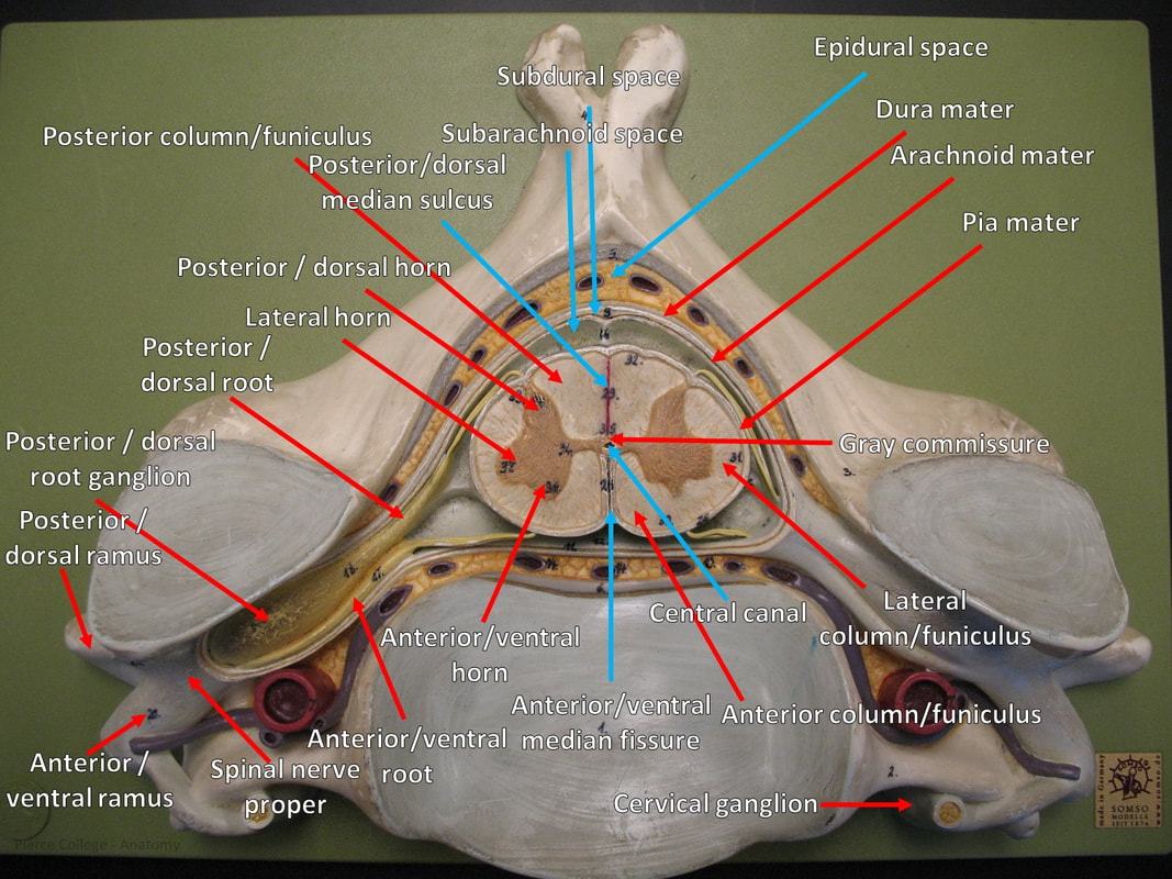
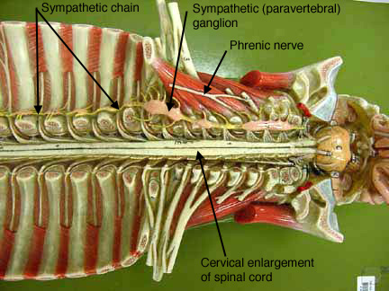
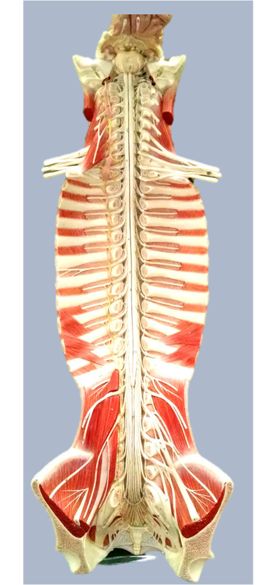
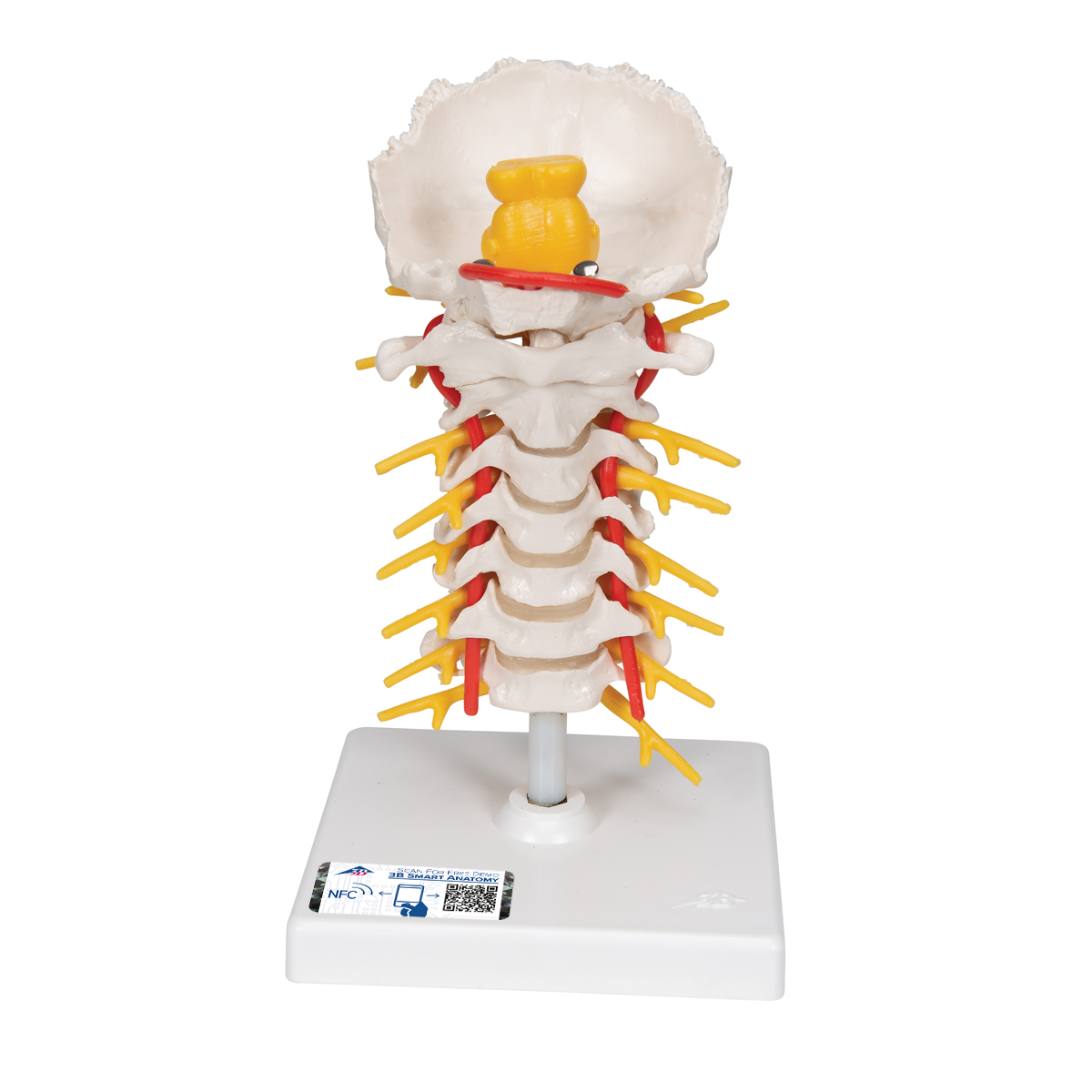
Spinal cord model with labels. Spine Anatomy, Diagram & Pictures | Body Maps - Healthline To facilitate this process, the spinal cord is divided into two kinds of pathways called tracts. Ascending tracts carry sensory input from the body to the brain, and descending tracts carry... Spinal Cord in the Spinal Canal (BS 31) · Anatomy models - SOMSO® Spinal Cord in the Spinal Canal. Seen from the ventral side, natural size, in SOMSO-PLAST®. The model shows the brain stem and the spinal cord, as well as the nerve branches, up to the coccygeal plexus. On the left side, the sympathetic trunk with its connections to the central nervous system is shown. In one piece. Mounted on a green board. Spinal Cord sections labeled Diagram | Quizlet Start studying Spinal Cord sections labeled. Learn vocabulary, terms, and more with flashcards, games, and other study tools. SPINAL CORD MODEL Flashcards | Quizlet Objectives for Spinal Cord (fifth cervic…. 210: Chapter 11 Blended Skills and Critical Thinki…. 5th - Social Studies Review - ch7 notes and questi….
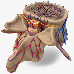
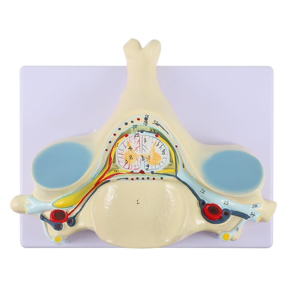
Spinal cord - Wikipedia The spinal cord is a long, thin, tubular structure made up of nervous tissue, which extends from the medulla oblongata in the brainstem to the lumbar region of the vertebral column (backbone). The backbone encloses the central canal of the spinal cord, which contains cerebrospinal fluid. The spinal cord | Human Anatomy and Physiology Lab (BSB 141 ... The spinal cord Information The spinal cord in cross-section has a central region of darker gray matter and the rest is lighter white matter. The gray matter is made up of neuroglia cells and neuron cell bodies. The white matter is made up of neuron axons, mostly but not all myelinated. PDF Anatomy & Physiology - TMCC Somso Model KS 4 Block model showing the skin with hair in different planes of section. I. Epidermis II. Corium (Dermis) III. Subcutis (Hypodermis) 1. External Horny Layer (Stratum corneum) 1a. Clear Layer (Stratum lucidum) -(KS 3 only) 2. Internal Hornless Germinative Zone (Stratum germinativum) 2a. Granular Layer (Stratum granulosum) 2b. Human Anatomy Torso Model Labeled Human Anatomy Torso Model Labeled Torso spinal cord human body posterior key. Organs torso omentum mesentery collin cauda equina gallbladder zaid 있는 digestive pueppi daffodilcooper. Anatomie brust torso weibliche weiblichen oberkörper muskulösen drüsen Human Anatomy Torso Model Labeled
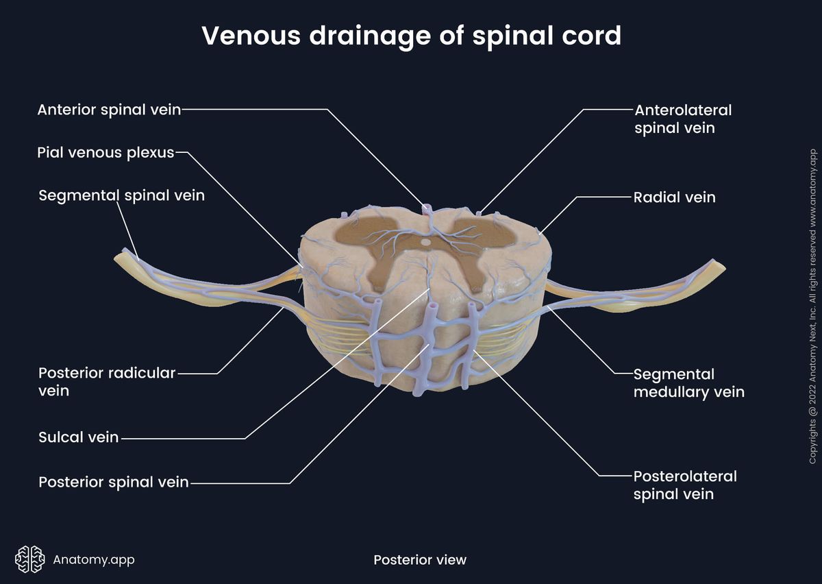
Labeled Brain Model Diagram | Science Trends The medial region of the posterior and anterior lobes function to control fine body movements, taking in input from the spinal cord as well as the auditory and visual systems of the brain. The lateral region of the cerebellum is the largest part of the cerebellum in humans. This region gets inputs from the cerebral cortex. Spinal cord: Anatomy, structure, tracts and function | Kenhub The spinal cord is made of gray and white matter just like other parts of the CNS. It shows four surfaces: anterior, posterior, and two lateral. They feature fissures (anterior) and sulci (anterolateral, posterolateral, and posterior). The gray matter is the butterfly-shaped central part of the spinal cord and is comprised of neuronal cell bodies. Patients with chronic pain used less sick leave after spinal ... Aug 31, 2022 · People with long-term neuropathic pain took significantly fewer sick days from work after treatment with spinal cord stimulation, according to a study by researchers at Karolinska Institutet ... Spinal Cord Diagram with Detailed Illustrations and Clear Labels Spinal Cord Diagram The spinal cord is one of the most important structures in the human body. It is the most important structure for any vertebrate. Anatomically, the spinal cord is made up of nervous tissue and is integrated into the spinal column of the backbone. Main Article: Spinal Cord - Anatomy, Structure, Function, and Spinal Cord Nerves
Lab - Spinal Cord: MAH-Summer 2019-Anatomy and Physiology I
Nervous System Models | Spinal Cord with Nerves Models Deluxe Spinal Cord Model (0165-00) Item # DGA65. $369.00 $339.00. Add to cart. Kyoto Kagaku Full-Figure Nervous System Model. Item # KK-A25. Add to cart. Kyoto Kagaku Nerves and Vessels of Arm Model. Item # KK-A144. Add to cart. Physiology of Nerves Series, 5 magnetics - illustrated metal board ...
BIOL 237 Class Notes - The Spinal Cord and Spinal Nerves
Nervous System Models - Labeled Brain and Spinal Cord - Pinterest Brain Model Labeled C Carmel Moore Nursing and Medical Health Science Spinal Cord Protection Just like the brain, the spinal cord is covered in both meninges and bone. The bone is what you'd typically think of as being the "spine". The meninges have the same names and... L Leslie Smart Physical Therapy Ear Anatomy Muscle Anatomy Facial Anatomy
Spinal Cord (Somso) - Pierce College - Anatomy
Learn the spinal cord with diagrams and quizzes | Kenhub The spinal cord, along with the brain, makes up the central nervous system (CNS). It is a long tubular structure comprised of nervous tissue, extending from the cervical to the lumbar region of the vertebral column. Just like other parts of the CNS, the spinal cord is comprised of white and gray matter. Spinal cord gray matter is the central ...
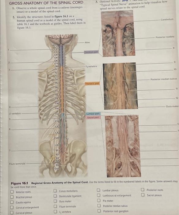
Solved 3. Optional Activity "Typical Spinal Nerve" animation ...
Spinal Cord Models - San Diego Mesa College Spinal Cord Models. photo for a larger view of the model. Click on Label for the labeled model. Back to Nervous System. Spinal Cord. (transverse section) Spinal Cord (close up) Spinal Cord. (longitudinal view)

Spinal cord: cross section model- pg 192 Flashcards | Quizlet
Lab Manual - Deep Back & Spinal Cord - Texas Tech University Health ... Consider the vertebral column as a whole and the structures which unite it: supraspinous ligament, ligamentum flavum, posterior longitudinal ligament, anterior longitudinal ligament (will be seen later), intervertebral disc. Identify intervertebral foramina, superior and inferior vertebral notches, and anterior and posterior sacral foramina. 2.

Spinal Anatomy | Vertebral Column
Can a human with a spinal cord injury walk and run ... Aug 15, 2022 · Mice with impaired spinal cord or motor nerve (left), recovery of motor function in mice with impaired nerve through stretchable artificial nerves (right). Credit: Seoul National University

Discectomy Demonstrator Model Simulator LXH - DDD Models
Spinal Cord Quiz: Cross-Sectional Anatomy | GetBodySmart Spinal Cord - Cross-Sectional Anatomy. Start Quiz. Want to learn faster? Look no further than these interactive, exam-style anatomy quizzes. Learn anatomy faster and remember everything you learn. Start Now. Related Articles. Parts of the Brain Quiz. Test your knowledge with the parts of the brain and their functions in a fun and interactive ...

Spinal Cord Model 5 times Life Size – GTSimulators.com
Spinal Cord Model.mov - YouTube Anatomy of Cross Section of Spinal Cord including spinal nerve, dorsal root ganglion, white matter, gray matter, central canal, dorsal gray horn, ventral gra...

NS_long_superior.jpg
Novel stochastic framework for automatic segmentation of ... Animal studies showed that spinal cord transection reduced muscle mass of hind-limb extensors between 20% and 40% in one month [2–4]. Individuals with chronic SCI also showed cross-sectional area of the whole thigh, knee extensors and plantar flexors that were about 30% smaller compared to non-disabled individuals [ 5 , 6 ].

Anatomical Model: Spinal Cord, No. 3
Amazon.com: spinal cord model 590 $14599 Get it as soon as Fri, May 6 FREE Shipping Only 8 left in stock - order soon. New-Horizon Scientific Spinal Cord Model,Skeleton Model -34" Life Size Spinal Column Model with Vertebrae, Nerves, Arteries, Lumbar Column, and Male Pelvis, Includes Stand (Good After-Sales) 10 Currently unavailable.
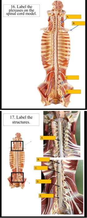
Solved 16. Label the plexuses on the spinal cord model. 17 ...
spinal cord anatomy, labeling spinal model Quiz - PurposeGames.com This is an online quiz called spinal cord anatomy, labeling spinal model There is a printable worksheet available for download here so you can take the quiz with pen and paper. Your Skills & Rank Total Points 0 Get started! Today's Rank -- 0 Today 's Points One of us! Game Points 16 You need to get 100% to score the 16 points available Actions
Spinal cord transverse section coverings label - Buy Royalty ...
spinal cord anatomy, labeling spinal model - Printable - PurposeGames.com About this Worksheet. This is a free printable worksheet in PDF format and holds a printable version of the quiz spinal cord anatomy, labeling spinal model.By printing out this quiz and taking it with pen and paper creates for a good variation to only playing it online.

anterior view of spinal cord - labeled (doesn't show ...
Axis Scientific Spine Model, 34" Life Size Spinal Cord Model with ... Axis Scientific Spine Model, 34" Life Size Spinal Cord Model with Vertebrae, Nerves, Arteries, Lumbar Column, and Male Pelvis, Includes Stand, Detailed Product Manual and Worry Free 3 Year Warranty ... Full Color Spine Model Study Guide . Includes 26 labeled parts! Read more. Read more. Read more. Brief content visible, double tap to read full ...

LS Spinal cord model Diagram | Quizlet
Spinal cord | Encyclopedia | Anatomy.app | Learn anatomy | 3D models ... Spinal cord segments and enlargements Like the vertebral column, the spinal cord anatomically can be divided into five parts: cervical, thoracic, lumbar, sacral and coccygeal. All parts correspond to the respective vertebral regions. The spinal cord is also arranged into segments. One segment corresponds to one pair of spinal nerves.
SCB209 - Lab1 - Natural Sciences Open Educational Resources
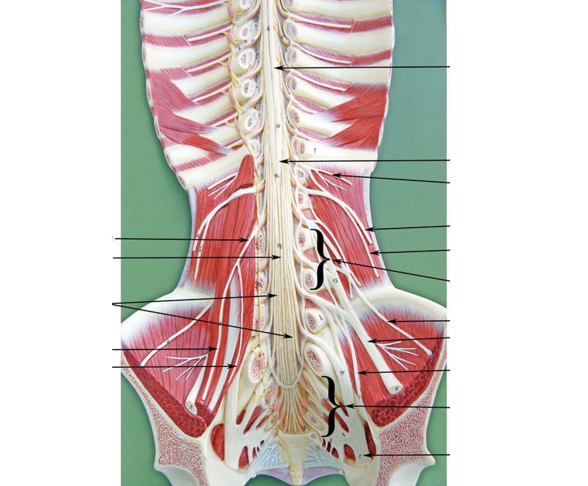
Solved 1. Label this picture of the model: Spinal cord and | Chegg.com 1. Label this picture of the model: Spinal cord and spinal column wall mount • Label: use the free drawing tool/brackets and arrows to Identify the following: • conus medullaris • cauda equina . filum terminale CU ; Question: 1. Label this picture of the model: Spinal cord and spinal column wall mount • Label: use the free drawing tool ...
Spinal Cord Models
How to tighten loose skin: 6 tips - Medical News Today Oct 03, 2019 · The effect of oral collagen peptide supplementation on skin moisture and the dermal collagen network: Evidence from an ex vivo model and randomized, placebo‐controlled clinical trials. https ...

154 Spinal Cord Labeled Stock Photos, Pictures & Royalty-Free ...
Anatomy of the spinal cord - e-Anatomy - IMAIOS This atlas of human anatomy describes the spinal cord through 18 anatomical diagrams with 270 anatomical structures labeled. It was designed particularly for physiotherapists, osteopaths, rheumatologists, neurosurgeons, orthopedic surgeons and general practitioners, especially for the study and understanding of medullary diseases.
3D Spine Models | TurboSquid
PDF Anatomy and Physiology of the Spinal Cord The spinal cord is a bundle of spinal nerves wrapped together. The spinal nerves enter and exit the spinal cord through small spaces between the vertebrae. The blood vessels which carry oxygen to the spinal cord also use these spaces. You have 8 pairs of cervical nerves, 12 thoracic, 5 lumbar and 6 sacral.
Lab - Spinal Cord: MAH-Summer 2019-Anatomy and Physiology I
Duke Neurosciences - Lab 2: Spinal Cord & Brainstem: Surface and ... Challenge 3.1—internal anatomy of the spinal cord. With reference to Figure 2.6, 2.7, and 2.8 and the chart below, carefully inspect the internal features of the spinal cord that are present in each segment, as well as those that are different (or present in only in one segment). To complete this challenge, spend some time browsing the spinal cord sections in Sylvius4, and find each of the ...

Lab: Spinal Cord Model Flashcards | Quizlet
Spinal Cord Injury | Types of Spinal Cord Injuries ... 88 percent of spinal cord injury survivors who were single at the time of the accident are single five years later, compared to 65 percent in the general population. Two-thirds of sports-related spinal cord injuries are from diving, making it the most dangerous sport for the brain and spinal cord.

Figure 2.1 from Fabrication of Gold-Platinum and Gold-Carbon ...

Nervous - Spinal Cord | Chandler Physical Therapy

Spinal cord Human vertebral column Cross section Nervous ...

NERVOUS SYSTEM ANATOMY: Cross section anatomy spinal cord

Spinal Cord – Torso – Human Body Help

Longitudinal Spinal Cord Model Diagram | Quizlet
Biology 2404 A&P Basics
Solved] Please help me labelling the parts of the spinal cord ...
Spinal Cord Models

Global-Dental Human Thoracic Vertebra Spinal Cord Spinal Nerve Sympathetic Trunk Magnifying Model
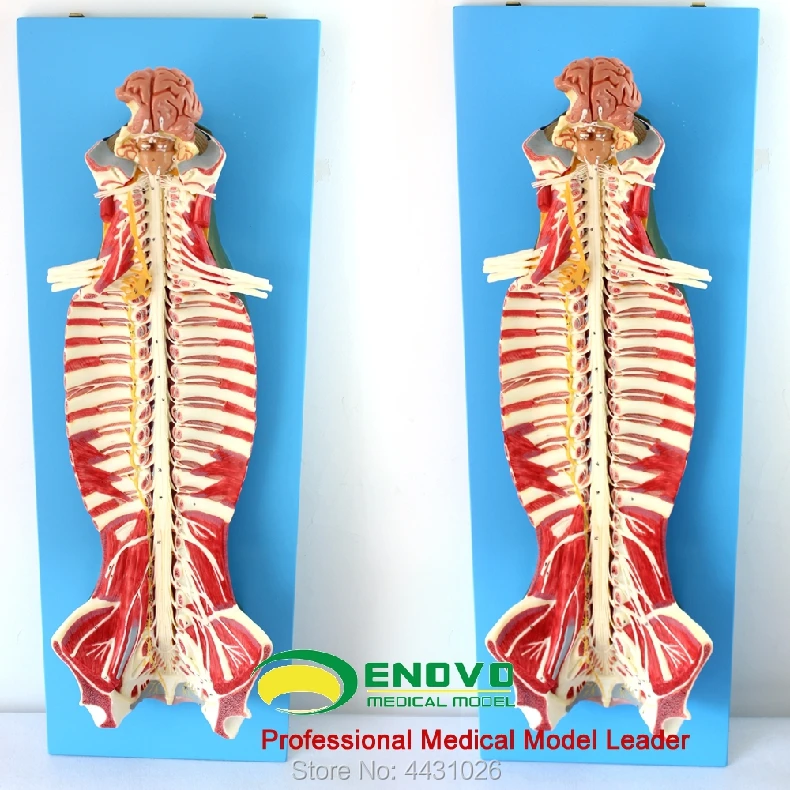
ENOVO Anatomy of spinal cord spinal nerve
15.5: Laboratory Activities and Assignment - Biology LibreTexts

spinal cord anatomy, labeling spinal model Quiz

Interactive Transverse Spinal Cord Labeling Quiz

Deluxe Spinal Cord | Denoyer-Geppert – Denoyer-Geppert ...

Anatomical Model: Spinal Cord, No. 3
Anatomical Teaching Models | Plastic Vertebrae Model ...
Spinal cord | Encyclopedia | Anatomy.app | Learn anatomy | 3D ...

Spinal Cord Diagram with Detailed Illustrations and Clear Labels
Spine Model

Spinal Cord Model.mov - YouTube
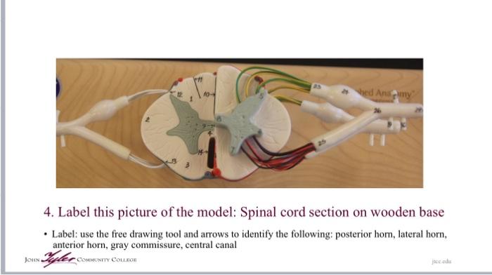
Solved hed Animy 4. Label this picture of the model: Spinal ...
Post a Comment for "42 spinal cord model with labels"