41 brain mri with labels
Brain MRI Segmentation Using FCM (Labeling) - Stack Overflow This can be accomplished with label2rgb as the documentation suggests here. I would probably use this form: RGB = label2rgb (L, map) Where map is a colormap. If you pass the same map to each slice's call to label2rgb the labels will be returned with the same colors. This can also be implemented relatively easily. › en › e-AnatomyCross-sectional anatomy of the brain - e-Anatomy - IMAIOS Apr 15, 2022 · Axial MRI Atlas of the Brain. Free online atlas with a comprehensive series of T1, contrast-enhanced T1, T2, T2*, FLAIR, Diffusion -weighted axial images from a normal humain brain. Scroll through the images with detailed labeling using our interactive interface. Perfect for clinicians, radiologists and residents reading brain MRI studies.
Deep learning to automate the labelling of head MRI datasets for ... manually labelling mri scans appears to be particularly laborious due to (1) the superior soft-tissue contrast of mri which enables more refined diagnoses compared with other imaging modalities such as computed tomography; and (2) the use of multiple, complementary imaging sequences so that a larger number of images must be scrutinised per …

Brain mri with labels
CaseStacks.com - MRI Brain Anatomy Labeled scrollable brain MRI covering anatomy with a level of detail appropriate for medical students. Show/Hide Labels. MRI Brain Anatomy. Back to Anatomy Overview. ... Labelled radiographs and CT/MRI series teaching anatomy with a level of detail appropriate for medical students and junior residents. Pelvis. Pelvic MRI anatomy › AANLIB › casesHarvard University Show labels Show list All modalities to: MR-T1 MR-T2 FDG T1/FDG T2/FDG UCLA Brain Mapping Center - ICBM Template To view both the structural MRI and the labels launch the program typing Display icbm_template.mnc -label icbm_labels_corrected.mnc. The opacity of the labels can be set in the Colour Coding menu. The number of each label appears at the bottom left of the orthogonal views window.
Brain mri with labels. Deep learning from MRI-derived labels enables automatic brain tissue ... Deep learning from MRI-derived labels enables automatic brain tissue classification on human brain CT Neuroimage. 2021 Dec 1;244:118606. doi: 10.1016/j ... Our proposed model predicted brain tissue classes accurately from unseen CT images (Dice coefficients of 0.79, 0.82, 0.75, 0.93 and 0.98 for GM, WM, CSF, brain volume and ICV, respectively). ... Arterial spin labeling MRI: clinical applications in the brain Visualization of cerebral blood flow (CBF) has become an important part of neuroimaging for a wide range of diseases. Arterial spin labeling (ASL) perfusion magnetic resonance imaging (MRI) sequences are increasingly being used to provide MR-based CBF quantification without the need for contrast administration, and can be obtained in conjunction with a structural MRI study. Brain lobes - annotated MRI | Radiology Case | Radiopaedia.org Debowski, M. Brain lobes - annotated MRI. Case study, Radiopaedia.org. (accessed on 08 Oct 2022) Brain MRI Dataset | Kaggle Kaggle is the world's largest data science community with powerful tools and resources to help you achieve your data science goals.
Brain MRI: How to read MRI brain scan | Kenhub MRI is the most sensitive imaging method when it comes to examining the structure of the brain and spinal cord. It works by exciting the tissue hydrogen protons, which in turn emit electromagnetic signals back to the MRI machine. The MRI machine detects their intensity and translates it into a gray-scale MRI image. Functional MRI of the Brain > Fact Sheets > Yale Medicine The exercises increase activity in specific parts of the brain, increasing blood flow and oxygen to them. This activity lights up on the images created by the scanner, giving doctors a visible record of an exact map of the patient's brain. A normal MRI of the brain can last between 20 to 30 minutes, while the fMRI lasts between 40 to 55 minutes. Magnetic Resonance Imaging (MRI) of the Spine and Brain Magnetic resonance imaging (MRI) is a diagnostic procedure that uses a combination of a large magnet, radiofrequencies, and a computer to produce detailed images of organs and structures within the body. Unlike X-rays or computed tomography (CT scans), MRI does not use ionizing radiation. Some MRI machines look like narrow tunnels, while others ... Atlas of BRAIN MRI - W-Radiology Brain magnetic resonance imaging (MRI) is a common medical imaging method that allows clinicians to examine the brain's anatomy (1). It uses a magnetic field and radio waves to produce detailed images of the brain and the brainstem to detect various conditions (2).
Frontiers | 101 Labeled Brain Images and a Consistent Human Cortical ... Labeled anatomical subdivisions of the brain enable one to quantify and report brain imaging data within brain regions, which is routinely done for functional, diffusion, and structural magnetic resonance images (f/d/MRI) and positron emission tomography data. brain anatomy | MRI coronal brain anatomy | free MRI cross sectional ... ELBOW AXIAL. WRIST AXIAL. WRIST CORONAL. KNEE CORONAL. KNEE SAGITTAL. ARTERIES UPPER LEG. ARTERIES LOWER LEG. This MRI brain coronal cross sectional anatomy tool is absolutely free to use. Use the mouse scroll wheel to move the images up and down alternatively use the tiny arrows (>>) on both side of the image to move the images. corner.bigblueinteractive.com › indexThe Corner Forum - New York Giants Fans Discussion Board ... Big Blue Interactive's Corner Forum is one of the premiere New York Giants fan-run message boards. Join the discussion about your favorite team! NITRC: Manually Labeled MRI Brain Scan Database: Tool/Resource Info Manually Labeled MRI Brain Scan Database Visit Website Image 1 of 3 Click for more. This is a continuously growing and improving database of high-quality neuroanatomically labeled MRI brain scans, created not by an algorithm, but by neuroanatomical experts. All results are checked and corrected.
Cross sectional Anatomy of Brain on... - World Of Radiology ...
What Does a Brain MRI Show? • San Diego Health What does a brain MRI show? The answer is, unfortunately, not very. MRI scans (magnetic resonance imaging) have been around for decades, and the technology has been steadily improving. Today, a brain MRI test can identify whether or not a person has a stroke, or if the person has suffered a traumatic brain injury, or if the person is suffering ...

File:MRI brain sagittal section.jpg - Wikimedia Commons
MRI anatomy | free MRI axial brain anatomy - Mrimaster.com This MRI brain cross sectional anatomy tool is absolutely free to use. Use the mouse scroll wheel to move the images up and down alternatively use the tiny arrows (>>) on both side of the image to move the images.
CaseStacks.com - MRI Brain Anatomy
en.wikipedia.org › wiki › Spinal_cord_injurySpinal cord injury - Wikipedia CT gives greater detail than X-rays, but exposes the patient to more radiation, and it still does not give images of the spinal cord or ligaments; MRI shows body structures in the greatest detail. Thus it is the standard for anyone who has neurological deficits found in SCI or is thought to have an unstable spinal column injury.

MRI anatomy | free MRI axial brain anatomy
Labeled MRI Brain Scans - Neuromorphometrics We can also label scans that you provide and we are very interested in labeling white matter anatomy as seen in diffusion-weighted MRI scans. If you want an aggregate version of our data, we can provide it as a probabilistic atlas. The cost to label a single scan is $2449 (USD).

Anatomy of the brain (MRI) | Mri brain, Anatomy, Mri
Researchers automate brain MRI image labeling, more than ... - ScienceDaily Researchers have automated brain MRI image labeling, needed to teach machine learning image recognition models, by deriving important labels from radiology reports and accurately assigning them to...
Magnetic resonance image (MRI) of a side view of the brain ...
Brain MRI Segmentation Using Pretrained 3-D U-Net Network Download Brain MRI and Label Data This example uses a subset of the CANDI data set [ 2] [ 3 ]. The subset consists of a brain MRI volume and the corresponding ground truth label volume for one patient. Both files are in the NIfTI file format. The total size of the data files is ~32 MB. Create a folder in which to store the data set.

Diagnostic usefulness of arterial spin labeling in MR ...
Head MRI: Purpose, Preparation, and Procedure - Healthline A head MRI is a useful tool for detecting a number of brain conditions, including: aneurysms, or bulging in the blood vessels of the brain; multiple sclerosis; spinal cord injuries; hydrocephalus ...
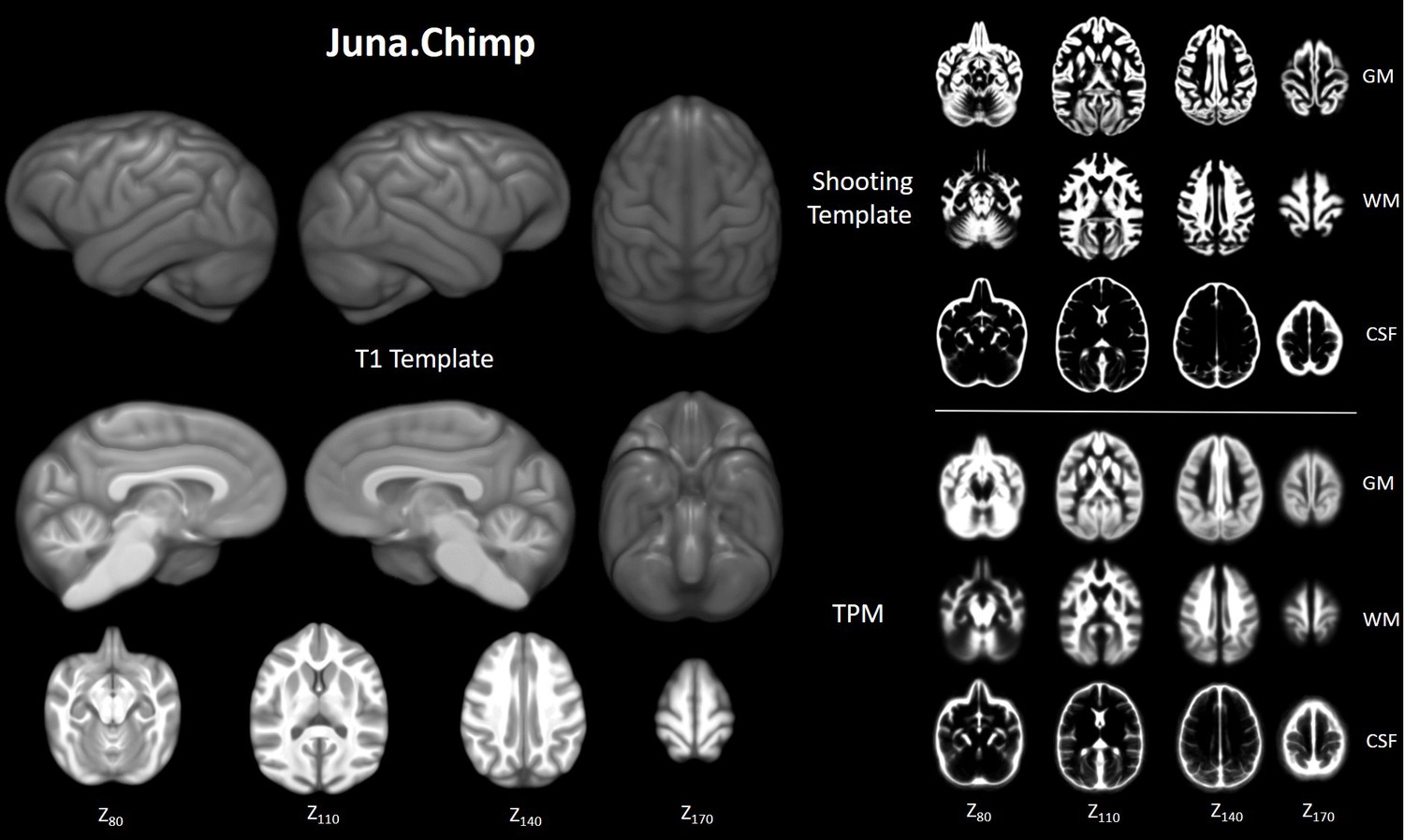
Chimpanzee brain morphometry utilizing standardized MRI ...
› home › brain,-spinal-cord,-andOverview of Spinal Cord Disorders - Brain, Spinal Cord, and ... The spinal cord Spinal Cord The spinal cord is a long, fragile tubelike structure that begins at the end of the brain stem and continues down almost to the bottom of the spine. The spinal cord consists of bundles of nerve... read more is the main pathway of communication between the brain and the rest of the body. It is a long, thick, fragile ...

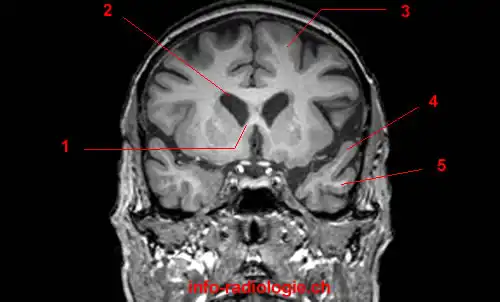
Label Each Part of the Brain Scan | MS in African Americans ...
Brain: Atlas of human anatomy with MRI - e-Anatomy - IMAIOS MRI Atlas of the Brain. This page presents a comprehensive series of labeled axial, sagittal and coronal images from a normal human brain magnetic resonance imaging exam. This MRI brain cross-sectional anatomy tool serves as a reference atlas to guide radiologists and researchers in the accurate identification of the brain structures.
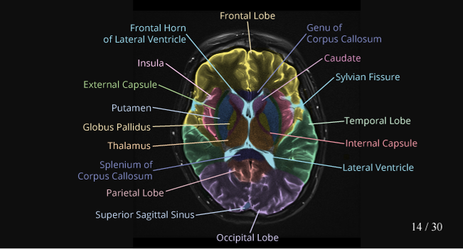
Approach to MRI brain | LearningNeurology.com
MRI Brain Animated Quiz - University of Minnesota MRI Brain Animated Quiz. Canine Brain MRI Anatomy Quiz. Sequentially click/tap: first the dot associated with a term; then, its corresponding target dot on the MRI image. If a line connection appears, your choice was correct! White Matter. Cerebral Cortex. Olfactory Bulb. Longitudinal Fissure.

MRI anatomy | free MRI axial brain anatomy
Labeled imaging anatomy cases | Radiology Reference Article ... This article lists a series of labeled imaging anatomy cases by body region and modality. Brain CT head: non-contrast axial CT head: non-contrast coronal CT head: non-contrast sagittal CT head: angiogram axial CT head: angiogram coronal CT...

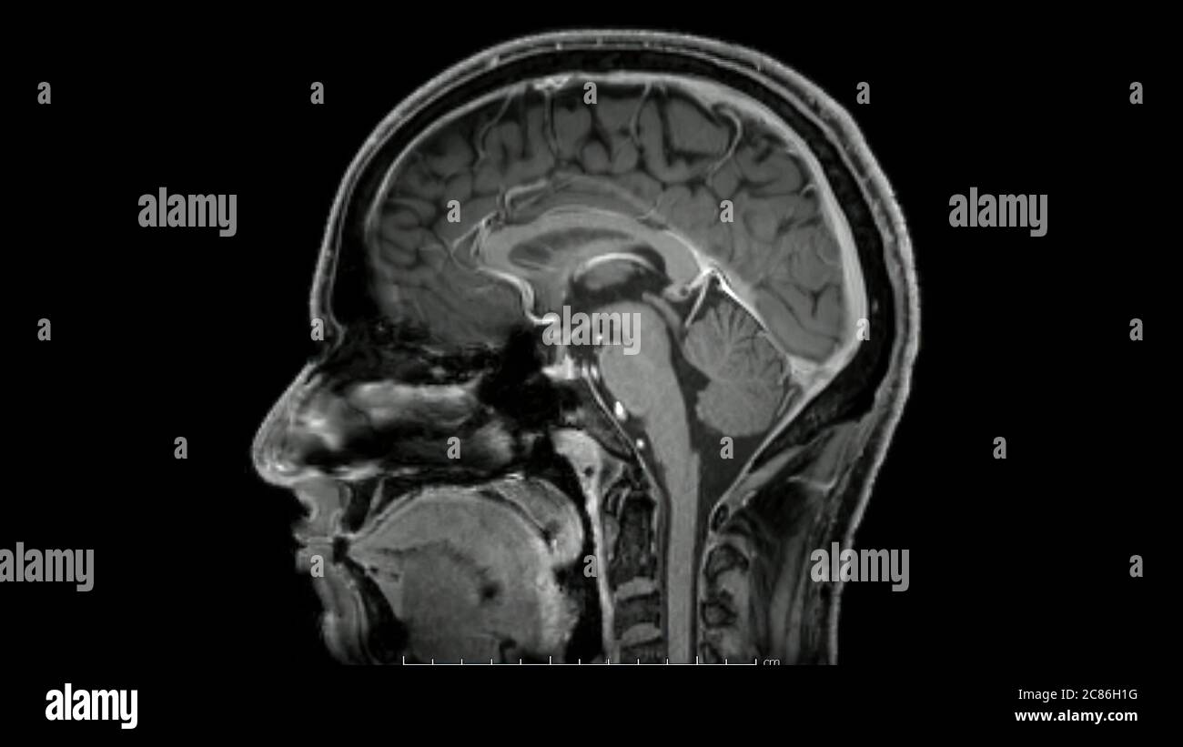
Normal anatomy of the brain on sagittal plane T1weighted ...
2,832 Labeled Brain Anatomy Images, Stock Photos & Vectors - Shutterstock Find Labeled brain anatomy stock images in HD and millions of other royalty-free stock photos, illustrations and vectors in the Shutterstock collection. Thousands of new, high-quality pictures added every day.

Brain: Atlas of human anatomy with MRI - e-Anatomy
101 Labeled Brain Images and a Consistent Human Cortical Labeling ... Labeled anatomical subdivisions of the brain enable one to quantify and report brain imaging data within brain regions, which is routinely done for functional, diffusion, and structural magnetic resonance images (f/d/MRI) and positron emission tomography data.
MRI identifies markers of atypical brain deve | EurekAlert!
pubmed.ncbi.nlm.nih.gov › 23878098Whole Brain Perfusion Measurements Using Arterial Spin ... The MB ASL technique is an effective method to evaluate whole brain perfusion because it minimizes the temporal spread of labeled spins across slices, resulting in more accurate perfusion measurements.
Frontiers | DeepNavNet: Automated Landmark Localization for ...
Brain MRI segmentation | Kaggle This dataset contains brain MR images together with manual FLAIR abnormality segmentation masks. The images were obtained from The Cancer Imaging Archive (TCIA). They correspond to 110 patients included in The Cancer Genome Atlas (TCGA) lower-grade glioma collection with at least fluid-attenuated inversion recovery (FLAIR) sequence and genomic ...
FDA Clears AI-Enabled Software for Streamlining Brain MRI ...
› cbica › brats-reg-challengeBrain Tumor Sequence Registration (BraTS-Reg) Challenge ... The data comprises of pairs of pre-operative baseline and follow-up MRI brain scans (each pair being of the same patient) diagnosed and treated for glioma. The exact multi-parametric MRI (mpMRI) sequences of each timepoint are i) native (T1) and ii) contrastenhancedT1-weighted (T1-CE), iii) T2-weighted and iv) T2Fluid Attenuated Inversion ...

Comparative Overview of Brain Perfusion Imaging Techniques ...
MRI Brain Atlas - University of Minnesota This web app Atlas is intended for veterinary students and radiologists seeking quick access to canine brain anatomy through a mobile device. Via a toggle button, either MRI images or approximately comparable Brain Transection images may be viewed with or without labels. Navigation & Labels.

MRI anatomy | free MRI axial brain anatomy
UCLA Brain Mapping Center - ICBM Template To view both the structural MRI and the labels launch the program typing Display icbm_template.mnc -label icbm_labels_corrected.mnc. The opacity of the labels can be set in the Colour Coding menu. The number of each label appears at the bottom left of the orthogonal views window.

Approach to MRI brain | LearningNeurology.com
› AANLIB › casesHarvard University Show labels Show list All modalities to: MR-T1 MR-T2 FDG T1/FDG T2/FDG

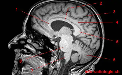
How to Read a MRI of Brain - Brain Anatomy MRI Explained in English
CaseStacks.com - MRI Brain Anatomy Labeled scrollable brain MRI covering anatomy with a level of detail appropriate for medical students. Show/Hide Labels. MRI Brain Anatomy. Back to Anatomy Overview. ... Labelled radiographs and CT/MRI series teaching anatomy with a level of detail appropriate for medical students and junior residents. Pelvis. Pelvic MRI anatomy

Automatic brain labeling via multi-atlas guided fully ...
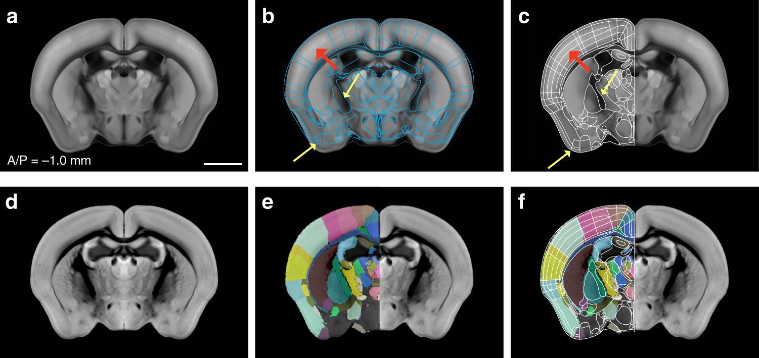
Enhanced and unified anatomical labeling for a common mouse ...
fMRI: Arterial Spin Labeling

volBrain: Automated MRI Brain volumetry system

Brain Anatomy and Images Brain
Brain MRI: How to read MRI brain scan | Kenhub

Neural Structure Quiz

Brain Anatomy MRI- Neuroradiology

volBrain: Automated MRI Brain volumetry system
Atlas of BRAIN MRI - W-Radiology
MRI Viewer on the App Store

Arterial Spin-Labeling Improves Detection of Intracranial ...

NeuroRad for iPad is a great app for medical professionals to ...
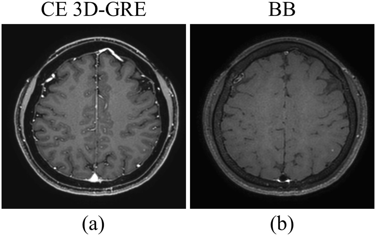
Deep-learned 3D black-blood imaging using automatic labelling ...

Brain imaging in MS,
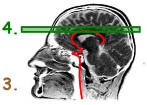
3 steps to optimize your MRI protocol

Labeled MRI Brain Scans

Magnetic Resonance Imaging (MRI): Brain - Connecticut Children's

Early postmortem brain MRI findings in COVID-19 non-survivors ...
Magnetic resonance images of the brain (MRI brain) sagittal ...

Brain scan, Mri scan, Psychology major
Atlas of BRAIN MRI - W-Radiology
Post a Comment for "41 brain mri with labels"